Anatomy Rib Cage : Anatomy Human Spine Torso And Rib Cage Buy Royalty Free 3d Model By Francescomilanese Francescomilanese 9624eb5 / The thoracic cage (rib cage) is the skeleton of the thoracic wall.
Anatomy Rib Cage : Anatomy Human Spine Torso And Rib Cage Buy Royalty Free 3d Model By Francescomilanese Francescomilanese 9624eb5 / The thoracic cage (rib cage) is the skeleton of the thoracic wall.. Womens body parts stomach 4 photos of the womens body parts stomach body diagram stomach, body parts digestive system, body parts in stomach area, body parts liver, body parts spleen, human body parts stomach, woman body organs, woman body parts found, stomach, body diagram stomach, body parts digestive system, body. Anatomy the rib cage is a bony structure found in the chest (thoracic cavity). As part of the bony thorax, the ribs protect the internal thoracic organs. 5 out of 5 stars (81) $ 16.71. At the chest, many rib bones connect to the sternum via costal cartilage,.
This image added by admin. Pain under the left rib cage can mean anything from a ruptured spleen, to heart trouble, to just needing to have a good fart. Thank you for visit anatomynote.com. Anatomy the rib cage is a bony structure found in the chest (thoracic cavity). Each pair is numbered based on their attachment to the sternum, a bony process at the front of the rib cage which serves as an anchor point.

It is made up of 12 pairs of ribs.
They are extremely light, but highly resilient; The thoracic cage (rib cage) is the skeleton of the thoracic wall. There is one last component of the axial skeleton we did not cover last lab: The thoracic cage (rib cage) is the skeletal framework of the thoracic wall, which encloses the thoracic cavity. We hope you can get the exact information. Thank you for visit anatomynote.com. See more ideas about anatomy reference, anatomy drawing, human anatomy. Rib 1 is also flattened horizontally. Rib bones are not classified as long bones.instead, anatomists classify the ribs as flat bones, and they are located within the axial skeleton.together with the sternum, thoracic vertebrae, and costal cartilages, the ribs form the thoracic cage, also called the bony thorax. Ribs 11 and 12 do not have necks or tubercles and the anterior tips of their bodies lack an articular surface. On the interior wall of the rib body is a channel, sulcus costae, with blood vessels and nerves. Anatomy the rib cage is a bony structure found in the chest (thoracic cavity). They articulate with the vertebral column posteriorly, and terminate anteriorly as cartilage (known as costal cartilage).
Related posts of muscle anatomy rib cage anatomy muscle cell. The ribs are curved, flat bones which form the majority of the thoracic cage. Ribs 11 and 12 do not have necks or tubercles and the anterior tips of their bodies lack an articular surface. The ribs are a set of twelve paired bones which form the protective 'cage' of the thorax. Anatomy muscle cell 12 photos of the anatomy muscle cell anatomy muscle cell, anatomy muscle cell.

5 out of 5 stars (81) $ 16.71.
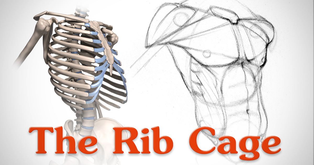
In this video, we explore:1) the anatomy of the sternum2) the anatomy and differences between the three classes of ribs3) the anatomy and differences between. The average skeleton contains 24 individual ribs, formed in 12. It has clear front, side, and back planes. At the chest, many rib bones connect to the sternum via costal cartilage,. Rib cage, in vertebrate anatomy, basketlike skeletal structure that forms the chest, or thorax, and is made up of the ribs and their corresponding attachments to the sternum (breastbone) and the vertebral column. Rib bones are not classified as long bones.instead, anatomists classify the ribs as flat bones, and they are located within the axial skeleton.together with the sternum, thoracic vertebrae, and costal cartilages, the ribs form the thoracic cage, also called the bony thorax. They articulate with the vertebral column posteriorly, and terminate anteriorly as cartilage (known as costal cartilage). Human rib cage anatomy 3d model. Anatomy of the rib cage the rib cage is a primarily protective structure, encircling the heart and lungs. The ribs are a set of twelve paired bones which form the protective 'cage' of the thorax. 5 out of 5 stars (81) $ 16.71. Contributing to their role in protecting the internal thoracic organs. This image added by admin.
A rib has a flat body, as you can see from the picture of the anatomy of the human rib cage. The ribs are a veritable collection of bone, muscle, and organs, most of which are fairly important for living and other useful functions. The thoracic cage takes the form of a domed bird cage with the horizontal bars formed by ribs and costal cartilages. Rib cage anatomy the rib cage, shaped in a mild cone shape and more flexible than most bone sets, is made up of varying elements such as the thoracic vertebra, 12 equally paired ribs, costal cartilage, and held together anteriorly by the sternum. Rib cage, in vertebrate anatomy, basketlike skeletal structure that forms the chest, or thorax, and is made up of the ribs and their corresponding attachments to the sternum (breastbone) and the vertebral column.
4 individual objects (spine portion, ribs, cartilages, sternum), sharing the same non overlapping uv layout map, material and pbr textures set.
4 individual objects (spine portion, ribs, cartilages, sternum), sharing the same non overlapping uv layout map, material and pbr textures set. Rib cage anatomy the rib cage, shaped in a mild cone shape and more flexible than most bone sets, is made up of varying elements such as the thoracic vertebra, 12 equally paired ribs, costal cartilage, and held together anteriorly by the sternum. Quads only geometries (no tris/ngons). The thoracic cage (rib cage) is the skeleton of the thoracic wall. 5 out of 5 stars (81) $ 16.71. It may occur after an obvious injury or without explanation. Anatomy muscle cell 12 photos of the anatomy muscle cell anatomy muscle cell, anatomy muscle cell. The top edge of the manubrium has a depression called the suprasternal or jugular notch. The upper edge is round and the lower sharp. Anatomy of the rib cage diagram in this image, you will find thoracic vertebrum, costochondral joint, costal cartilage, costal margin, costal arch, thoracic vertebrum, xiphoid process, xiphisternal joint, body, manubrial sternal joint, manubrium, the sternal notch in it. The bones of the rib cage are the sternum, the 12 thoracic vertebrae and the 12 pairs of ribs. As part of the bony thorax, the ribs protect the internal thoracic organs. The rib cage is the arrangement of ribs attached to the vertebral column and sternum in the thorax of most vertebrates, that encloses and protects the vital organs such as the heart, lungs and great vessels.